

-
Welcome to Tacoma World!
You are currently viewing as a guest! To get full-access, you need to register for a FREE account.
As a registered member, you’ll be able to:- Participate in all Tacoma discussion topics
- Communicate privately with other Tacoma owners from around the world
- Post your own photos in our Members Gallery
- Access all special features of the site
 WTB BAMF Thermoplastic Powder
 FS: Jack Accessories
 Cybermonday 2016
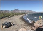 New to NorCal and to off roading.
 San Francisco "any places to go off roading"
Norcal Spotted and BS Thread
Discussion in 'Northern California' started by PreRunnerSeth, Sep 23, 2009.
Page 3361 of 8453
Page 3361 of 8453
Products Discussed in


